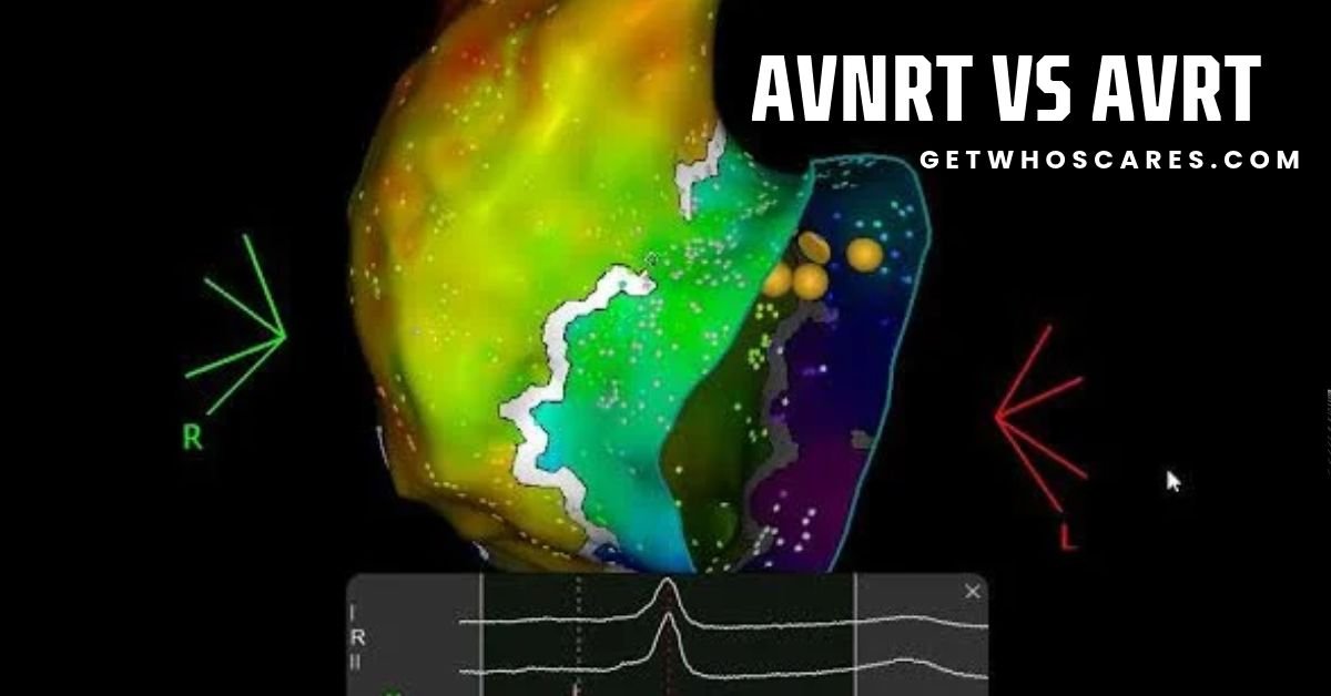
AVNRT vs AVRT: ECG Differences, Causes, Symptoms, and Treatment
AVNRT vs AVRT is a common topic in cardiology because both conditions are types of supraventricular tachycardia (SVT). They can cause a sudden, rapid heartbeat and may look very similar on an electrocardiogram (ECG or EKG). However, they have different electrical pathways inside the heart, and understanding these differences helps doctors identify the rhythm and choose the right treatment.
AVNRT stands for atrioventricular nodal reentrant tachycardia. AVRT stands for atrioventricular reentrant tachycardia. Both conditions involve an electrical signal traveling repeatedly through a circuit, causing the heart to beat faster than normal. The main difference is the location and structure of that circuit.
In AVNRT, the electrical circuit usually involves two pathways in or near the atrioventricular (AV) node. In AVRT, the circuit uses the normal electrical conduction system and an additional pathway connecting the atria and ventricles.
Both conditions can occur in people with otherwise healthy hearts. Symptoms may include a racing heartbeat, dizziness, shortness of breath, chest discomfort, or a feeling that the heart suddenly starts and stops beating rapidly.
This guide explains the AVNRT vs AVRT difference in simple language. It covers their full forms, causes, symptoms, ECG findings, RP intervals, treatment options, and long-term outlook. It also explains how doctors distinguish these rhythms from atrial tachycardia and other forms of SVT.
This article is for educational purposes. It does not replace medical advice, ECG interpretation by a qualified clinician, or emergency care.
What Is AVNRT?
AVNRT, or atrioventricular nodal reentrant tachycardia, is a fast heart rhythm caused by an electrical signal repeatedly traveling through a small circuit in or near the AV node.
The AV node is a group of specialized cells that helps control the electrical signal traveling from the upper chambers of the heart, called the atria, to the lower chambers, called the ventricles.
Normally, the electrical signal moves from the atria through the AV node and then into the ventricles. The AV node also slows the signal briefly, allowing the ventricles time to fill with blood before they contract.
In AVNRT, some people have two pathways with different electrical properties in the AV nodal region. These are commonly called the fast pathway and the slow pathway.
Under certain conditions, an early heartbeat can travel down one pathway and return through the other. This creates a repeating electrical loop known as reentry.
As the signal continues to circulate, the heart beats rapidly. The rhythm may begin suddenly and stop just as suddenly.
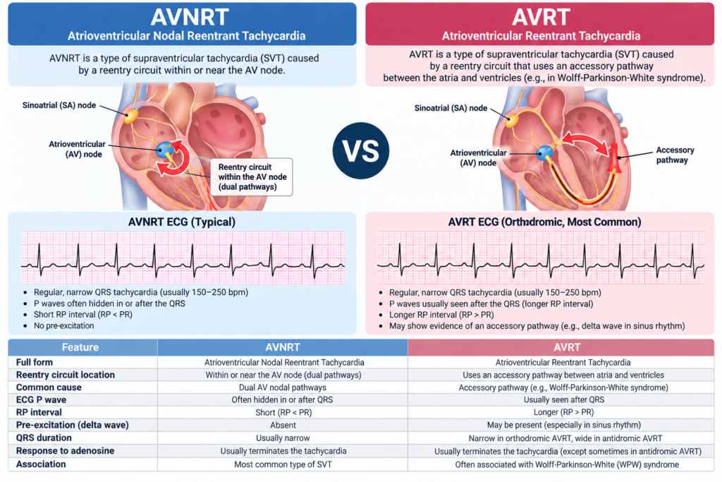
AVNRT meaning and medical definition
The term AVNRT has three main parts:
- Atrioventricular: relating to the atria and ventricles.
- Nodal: referring to the AV node region.
- Reentrant tachycardia: a rapid heart rhythm caused by an electrical signal repeatedly traveling around a circuit.
AVNRT is one of the most common forms of paroxysmal supraventricular tachycardia. Paroxysmal means that episodes begin and end suddenly.
A typical episode may produce a regular heartbeat of around 150 to 250 beats per minute, although the actual rate varies. The rhythm can occur in people without major structural heart disease.
What causes AVNRT?
AVNRT is usually related to differences in electrical conduction and recovery time within the AV nodal region. It is not necessarily caused by a blocked artery, a weak heart muscle, or a lifestyle problem.
An episode may begin after a premature atrial contraction, which is an early heartbeat originating in the atria. The early signal may find one pathway temporarily unable to conduct. It then travels through the other pathway and returns through tissue that has recovered, starting the repeating circuit.
Possible triggers include exercise, emotional stress, lack of sleep, dehydration, alcohol, caffeine, and stimulant use in some people. However, triggers vary, and many episodes occur without an obvious cause.
AVNRT may also occur in people who have no known family history of arrhythmia.
Typical and atypical AVNRT
There are different forms of AVNRT.
Typical AVNRT, often called slow-fast AVNRT, is the most common form. The signal usually travels forward through the slow pathway and returns through the fast pathway.
Atypical AVNRT is less common. It can involve different combinations of pathways, such as fast-slow or slow-slow conduction. These forms may produce a more visible delay between ventricular activation and the next atrial activation.
Also Read: Ligament vs Tendon: Key Differences, Functions, Injuries, and Treatment
This distinction matters because typical and atypical AVNRT can have different ECG appearances, especially when doctors assess the P wave and RP interval.
What Is AVRT?
AVRT stands for atrioventricular reentrant tachycardia. It is a fast heart rhythm caused by an electrical circuit that uses the AV node and an additional electrical connection between the atria and ventricles.
This additional connection is called an accessory pathway. It allows electrical signals to travel between the upper and lower chambers outside the usual AV nodal route.
During AVRT, the signal travels around a loop involving the normal conduction system and the accessory pathway. As the signal repeatedly completes the circuit, the heart beats rapidly.
AVRT is another important cause of paroxysmal supraventricular tachycardia. It is particularly associated with accessory pathways found in people with Wolff-Parkinson-White syndrome or those who have a concealed accessory pathway.
AVRT meaning and medical definition
The full form of AVRT is atrioventricular reentrant tachycardia. It is also described in medical literature as atrioventricular reciprocating tachycardia.
The word reciprocating refers to the electrical signal moving back and forth through a circuit. The term reentrant describes the signal returning to tissue that can conduct it again.
Unlike typical AVNRT, AVRT depends on an accessory pathway outside the AV node region as part of the reentry circuit.
What causes AVRT?
AVRT occurs when an accessory electrical pathway connects the atria and ventricles. This pathway is usually present from birth, although the rhythm problem may not become noticeable until later in life.
An accessory pathway can conduct electrical signals from the atria to the ventricles, from the ventricles to the atria, or in both directions.
Some pathways conduct only backward, from the ventricles to the atria. These are called concealed accessory pathways. They may cause AVRT even when a resting ECG does not show the classic signs of Wolff-Parkinson-White syndrome.
Other pathways conduct forward to the ventricles and may produce pre-excitation on a resting ECG.
An accessory pathway does not always cause symptoms. Some people have one without experiencing tachycardia, while others develop repeated episodes of rapid heartbeat.
AVRT and Wolff-Parkinson-White syndrome
Wolff-Parkinson-White (WPW) syndrome is an important condition associated with AVRT. It involves an accessory pathway that can allow electrical signals to bypass the normal delay in the AV node.
When an accessory pathway conducts forward during normal sinus rhythm, the ECG may show a short PR interval and a delta wave, which is a slurred beginning of the QRS complex.
However, not every person with AVRT has a visible delta wave. A concealed accessory pathway can produce AVRT without the typical resting ECG pattern of WPW.
It is important to distinguish having an accessory pathway from having an active episode of AVRT. An accessory pathway is an anatomical and electrical feature; AVRT is the tachycardia that occurs when a reentry circuit uses it.
AVNRT vs AVRT: Main Differences at a Glance
The most important difference between AVNRT and AVRT is the electrical circuit responsible for the fast rhythm.
| Feature | AVNRT | AVRT |
| Full form | Atrioventricular nodal reentrant tachycardia | Atrioventricular reentrant tachycardia |
| Main circuit | Usually involves pathways in or near the AV node | Uses the AV node and an accessory pathway |
| Accessory pathway required | No | Yes |
| Typical ECG during an episode | Often narrow QRS with a P wave hidden in or close to the QRS | Often narrow QRS in orthodromic AVRT, with a retrograde P wave after the QRS |
| P-wave appearance | May be hidden or appear as a pseudo-R’ in V1 or pseudo-S waves in inferior leads | Often visible shortly after the QRS in orthodromic AVRT |
| RP interval | Usually very short in typical AVNRT | Often short in orthodromic AVRT, but depends on the pathway and rhythm |
| Resting ECG between episodes | Often normal | May show pre-excitation if the accessory pathway conducts forward |
| WPW association | Not required | Important association |
| Symptoms | Sudden palpitations, dizziness, breathlessness, or chest discomfort | Similar symptoms |
| Acute treatment | Vagal maneuvers and, when appropriate, adenosine or other medical treatment | Similar treatment for appropriate stable rhythms, with important exceptions |
| Long-term treatment | Observation, medication, or catheter ablation | Observation, medication, or catheter ablation |
These are typical patterns, not absolute rules. A surface ECG may not clearly separate AVNRT from AVRT in every case. A cardiologist may need additional ECG recordings or an electrophysiology study to establish the diagnosis.
AVNRT vs AVRT ECG: How to Read the Differences
An ECG records the heart’s electrical activity. Doctors use the timing and shape of the electrical waves to identify the rhythm.
When comparing AVNRT vs AVRT on ECG, the main features include the QRS complex, P waves, RP interval, PR interval, and the relationship between atrial and ventricular activation.
A standard 12-lead ECG recorded during an episode is often more useful than a normal ECG recorded between episodes.
1. QRS complex
The QRS complex represents electrical activation of the ventricles.
Typical AVNRT usually produces a narrow QRS complex because the signal reaches the ventricles through the normal His-Purkinje conduction system.
Orthodromic AVRT also usually produces a narrow QRS complex because the signal travels forward through the AV node and normal conduction system to activate the ventricles.
Therefore, a narrow QRS complex alone cannot reliably distinguish AVNRT from orthodromic AVRT.
A wide QRS complex may occur with a pre-existing bundle branch block or another conduction problem. AVRT may also produce a wide QRS complex during antidromic conduction.
A wide-complex tachycardia requires careful medical assessment. If its cause is uncertain, clinicians must consider ventricular tachycardia rather than automatically assuming it is a form of SVT.
2. P waves
P waves represent electrical activation of the atria.
In typical AVNRT, atrial and ventricular activation occur very close together. The P wave may therefore be hidden inside the QRS complex.
Sometimes, the P wave creates a small extra deflection at the end of the QRS complex. In lead V1, this may appear as a pseudo-R’ wave. In the inferior leads II, III, and aVF, it may appear as a pseudo-S wave.
These findings can support a diagnosis of typical AVNRT, but their absence does not rule it out.
In orthodromic AVRT, the electrical signal activates the ventricles through the AV node and then returns to the atria through the accessory pathway. A retrograde P wave may therefore appear shortly after the QRS complex.
The P wave can be easier to identify in AVRT than in typical AVNRT. However, the difference is not always clear because some AVRT pathways produce very short intervals between ventricular and atrial activation.
3. PR interval
The PR interval measures the time from the beginning of atrial activation to the beginning of ventricular activation during a conducted heartbeat.
During a rapid rhythm, the P wave may be hidden or may not be easy to identify. This can make the PR interval difficult to measure.
In typical AVNRT, the atria and ventricles may activate almost at the same time, making the P wave difficult to separate from the QRS.
In orthodromic AVRT, the P wave usually follows the QRS during the tachycardia. The PR interval for that retrograde P wave is not measured in the same way as the interval between a normal P wave and its following QRS.
Doctors should therefore avoid using the PR interval alone to decide between AVNRT and AVRT.
4. RP interval
The RP interval is especially useful when comparing AVNRT vs AVRT ECG findings.
It measures the time from the beginning of a QRS complex to the beginning of the next visible retrograde P wave. In practice, clinicians interpret it alongside the PR interval and the full rhythm pattern.
A short RP tachycardia has a short interval between ventricular activation and the next visible atrial activation. Typical AVNRT and orthodromic AVRT commonly fall into this group.
In typical AVNRT, the RP interval is often extremely short because atrial activation occurs almost at the same time as ventricular activation.
In orthodromic AVRT, the RP interval may be longer because the electrical signal must travel from the ventricles back to the atria through the accessory pathway. However, a pathway located close to the AV node may produce a very short interval.
A long RP interval may suggest atypical AVNRT, certain forms of AVRT, or atrial tachycardia. It does not identify one diagnosis by itself.
The European Society of Cardiology explains that RP and PR relationships are useful parts of the differential diagnosis of supraventricular tachycardia, but additional findings may be needed to distinguish similar rhythms.
5. Resting ECG between episodes
The ECG between episodes can provide another important clue.
People with AVNRT often have a normal resting ECG when they are not experiencing tachycardia.
Also Read: Flautas vs. Taquitos: What’s the Difference Between These Mexican Foods?
People with an accessory pathway that conducts forward may show signs of pre-excitation, including a short PR interval and a delta wave. These findings can suggest WPW pattern.
However, a normal resting ECG does not exclude AVRT. A concealed accessory pathway may conduct only backward and leave no obvious sign of pre-excitation during normal sinus rhythm.
For this reason, a normal ECG between episodes should not be used as the only reason to rule out AVRT.
AVNRT vs AVRT Diagram: Understanding the Electrical Circuits
A simple text diagram can help explain the main difference between these rhythms.
AVNRT circuit
A typical AVNRT circuit uses two functional pathways in or near the AV node.
A simplified sequence is:
Early atrial beat → slow pathway → lower conduction system → fast pathway in the reverse direction → atrial tissue → repeating circuit.
The electrical signal keeps moving around the loop. Each cycle can activate the ventricles and produce another heartbeat.
The fast and slow pathways are functional conduction routes. They are not necessarily two separate accessory connections running between the atria and ventricles.
AVRT circuit
AVRT uses the normal AV conduction system and an accessory pathway.
In orthodromic AVRT, the sequence is:
Atria → AV node → His-Purkinje system → ventricles → accessory pathway → atria → repeating circuit.
In antidromic AVRT, the direction of forward conduction is reversed:
Atria → accessory pathway → ventricles → normal conduction system in the reverse direction → AV node region and atria → repeating circuit.
The exact circuit depends on the location and conduction properties of the accessory pathway.
The key point is that AVNRT usually depends on reentry in the AV nodal region, while AVRT depends on an accessory pathway connecting the atria and ventricles.
Orthodromic AVRT vs Antidromic AVRT
AVRT has two main forms, based on the direction in which the electrical signal reaches the ventricles.
What is orthodromic AVRT?
Orthodromic AVRT is the more common form of AVRT.
The electrical signal travels forward from the atria through the AV node and the normal conduction system to activate the ventricles. It then returns to the atria through the accessory pathway.
Because the ventricles are activated through the normal conduction system, the QRS complex is usually narrow unless another conduction problem is present.
Orthodromic AVRT ECG findings often include a regular, narrow-complex tachycardia with a retrograde P wave shortly after the QRS complex.
This appearance can resemble AVNRT, so the P-wave timing and other clinical findings are important.
What is antidromic AVRT?
Antidromic AVRT is less common.
In this rhythm, the electrical signal travels forward from the atria to the ventricles through the accessory pathway. The ventricles are therefore activated outside the normal rapid conduction system, which commonly produces a wide QRS complex.
The signal then returns toward the atria through the normal conduction system or, in some cases, another accessory pathway.
An antidromic AVRT ECG may look similar to ventricular tachycardia because both can produce a rapid rhythm with wide QRS complexes.
This is a clinically important distinction. A wide-complex rhythm should not be assumed to be antidromic AVRT without appropriate evidence.
Antidromic AVRT vs ventricular tachycardia
Ventricular tachycardia, or VT, starts in the ventricles. Antidromic AVRT is a reentrant rhythm that involves an accessory pathway and the normal conduction system.
Both may produce wide QRS complexes, but their electrical mechanisms are different.
Doctors may use the patient’s history, the ECG during the episode, evidence of an accessory pathway, and specialized electrophysiology testing to distinguish them.
When the cause of a wide-complex tachycardia is uncertain, emergency clinicians generally manage it as potentially serious ventricular tachycardia until a safer diagnosis is established.
Antidromic AVRT and WPW
Antidromic AVRT can occur in people with an accessory pathway associated with WPW.
The wide QRS complex reflects ventricular activation through the accessory pathway rather than the usual rapid conduction system.
Treatment choices differ from those for a typical narrow-complex SVT. Some medicines that block the AV node can be dangerous in pre-excited atrial fibrillation, so the rhythm mechanism must be identified before selecting treatment.
Adenosine may be used in selected, carefully assessed regular tachycardias, but it is not a medicine for unsupervised self-treatment. Its use in wide-complex rhythms requires particular caution and appropriate monitoring.
AVNRT vs AVRT vs Atrial Tachycardia
AVNRT and AVRT are both reentrant rhythms that usually depend on the AV node as part of the circuit. Atrial tachycardia is different because the rhythm starts in an area of atrial tissue that generates rapid electrical signals.
The three conditions may produce a fast heartbeat, but their electrical mechanisms differ.
| Feature | AVNRT | AVRT | Atrial tachycardia |
| Main source of rhythm | Reentry in or near the AV node | Reentry using an accessory pathway | An area of atrial tissue |
| Role of AV node | Part of the circuit | Usually part of the circuit | May conduct signals but is not necessarily part of the atrial rhythm mechanism |
| P waves | Often hidden or very close to the QRS in typical AVNRT | Often appear after the QRS in orthodromic AVRT | Often visible before the QRS, depending on the location and conduction |
| QRS complex | Usually narrow | Usually narrow in orthodromic AVRT; often wide in antidromic AVRT | Usually narrow if ventricular conduction is normal |
| Effect of AV nodal block | Usually interrupts the circuit | Usually interrupts the circuit | May slow ventricular conduction without stopping the atrial rhythm |
| Resting ECG clues | Often normal | May show pre-excitation | Depends on the specific atrial rhythm |
How doctors tell them apart
Doctors assess whether the atrial activity continues when conduction through the AV node is temporarily blocked.
AVNRT and AVRT usually require conduction through the AV node as part of their circuits. Blocking that conduction can interrupt the rhythm.
Atrial tachycardia can continue even when the AV node temporarily stops conducting signals to the ventricles. In such cases, the ECG may show atrial activity without a corresponding QRS complex.
However, these responses are not perfect diagnostic tests. Some atrial tachycardias can stop with certain interventions, and the findings must be interpreted in context.
Symptoms of AVNRT and AVRT
The symptoms of AVNRT and AVRT overlap because both can cause a rapid heart rhythm.
Common symptoms include:
- Sudden, noticeable heart racing or pounding.
- A regular, fast pulse.
- Fluttering or pounding in the chest.
- Dizziness or lightheadedness.
- Shortness of breath.
- Chest pressure or discomfort.
- Weakness or unusual tiredness.
- Anxiety or a sense of alarm during an episode.
- In some cases, fainting or near-fainting.
Some people describe a strong pulsing sensation in the neck. In AVNRT, this may occur because the atria and ventricles contract close together, causing pressure changes in the neck veins. This is sometimes called the frog sign.
Symptoms alone cannot reliably distinguish AVNRT from AVRT. A person with either condition may have mild symptoms or severe discomfort, depending on the heart rate, episode duration, underlying health, and other factors.
Seek emergency medical help for a rapid heartbeat accompanied by fainting, severe chest pain, severe shortness of breath, confusion, or signs of collapse. Do not wait for the rhythm to stop on its own if severe symptoms occur.
How Are AVNRT and AVRT Diagnosed?
A diagnosis usually combines the patient’s medical history, a physical examination, ECG recordings, and, when necessary, specialized testing.
1. Twelve-lead ECG
A 12-lead ECG records electrical activity from several views of the heart.
The most useful recording is often one taken during the rapid heartbeat. It can show the rate, regularity, QRS width, P-wave position, and RP relationship.
A recording made after the episode ends may be normal, especially in AVNRT or AVRT caused by a concealed pathway.
2. Holter monitor or event monitor
If episodes occur only occasionally, a short ECG may not capture them.
A Holter monitor records the heart rhythm continuously for a set period, often one or two days. An event monitor or another ambulatory ECG device may record the rhythm over a longer period.
The appropriate device depends on how often symptoms occur and how likely they are to be captured.
3. Wearable devices
Some smartwatches and portable ECG devices can record heart rhythm information. These recordings may help document an episode, but they do not always provide enough information to distinguish AVNRT from AVRT.
A wearable device should not replace a medical evaluation, especially when symptoms are new, severe, or associated with fainting or chest pain.
4. Electrophysiology study
An electrophysiology study, or EP study, is a specialized procedure used to examine the electrical system inside the heart.
Doctors guide thin catheters with electrical sensors through blood vessels into the heart. They record electrical signals and may use controlled pacing to reproduce the abnormal rhythm.
An EP study can help identify the reentry circuit, distinguish AVNRT from AVRT, locate an accessory pathway, and guide catheter ablation when appropriate.
This test is especially useful when the diagnosis remains uncertain or when a person is considering a potentially curative procedure.
Treatment of AVNRT vs AVRT
Treatment depends on the type of rhythm, how often episodes occur, the severity of symptoms, other medical conditions, and the person’s preferences.
Options include observation, techniques that stimulate the vagus nerve, medicines, and catheter ablation.
1. Vagal maneuvers
Vagal maneuvers are techniques that increase the activity of the vagus nerve and can temporarily slow conduction through the AV node.
A clinician may teach an appropriate technique, such as a modified Valsalva maneuver, for selected episodes of a regular SVT.
These techniques can stop many episodes of AVNRT and orthodromic AVRT because the AV node is part of their reentry circuits.
They do not work for every episode and should not delay emergency care when severe symptoms are present. Carotid sinus massage should not be attempted by an untrained person.
2. Adenosine
Adenosine is a short-acting medicine used in monitored medical settings to interrupt certain regular tachycardias that depend on the AV node.
It can terminate many episodes of AVNRT and orthodromic AVRT.
Adenosine may also help reveal the underlying atrial activity by temporarily blocking AV nodal conduction. This can help clinicians distinguish a reentrant rhythm from some other forms of tachycardia.
It may cause a brief feeling of chest pressure, flushing, breathlessness, or an unusual sensation. These effects usually pass quickly, but administration requires appropriate clinical monitoring.
Adenosine is not appropriate for every fast rhythm. In particular, an irregular or pre-excited wide-complex rhythm requires a different approach and careful assessment.
3. Other medicines
Depending on the rhythm and the patient’s condition, clinicians may use medicines such as beta blockers or certain calcium channel blockers to reduce episodes or slow conduction through the AV node.
These medicines can be useful for selected patients, but they are not suitable for everyone. The choice depends on factors such as blood pressure, heart function, other medicines, and the exact rhythm diagnosis.
Medicines that slow the AV node can be harmful in some forms of pre-excited atrial fibrillation associated with an accessory pathway. This is one reason accurate rhythm assessment is essential.
4. Synchronized cardioversion
Synchronized electrical cardioversion may be needed when a tachycardia causes serious symptoms or unstable blood circulation, or when appropriate medication does not work.
The procedure delivers a controlled electrical shock timed with the heart’s electrical cycle to restore a safer rhythm.
Emergency treatment decisions depend on the patient’s condition and the type of rhythm. Severe symptoms require immediate medical assessment rather than attempts at home treatment.
5. Catheter ablation
Catheter ablation is a treatment that targets the tissue responsible for the abnormal electrical circuit.
For AVNRT, the procedure usually modifies the slow pathway in the AV nodal region. The goal is to prevent the circuit from continuing while preserving normal AV conduction.
For AVRT, the procedure targets the accessory pathway that supports the reentry circuit.
Ablation is often highly effective for both conditions and may provide long-term freedom from recurrent episodes. However, success rates and risks differ based on the rhythm mechanism, pathway location, patient characteristics, and the experience of the treatment center.
Potential complications include bleeding, blood vessel injury, infection, damage to nearby cardiac structures, and, rarely, damage to normal AV conduction requiring a pacemaker. The risks should be discussed with an electrophysiologist before the procedure.
For many people with frequent or disruptive episodes, ablation can be an important alternative to taking long-term medication.
Is AVNRT More Dangerous Than AVRT?
Neither condition is automatically more dangerous in every person. The risk depends on the exact rhythm, the accessory pathway’s properties, associated heart disease, and the symptoms experienced during an episode.
AVNRT is often well tolerated in people with otherwise healthy hearts, although a very rapid rhythm can cause significant discomfort, dizziness, or reduced blood pressure.
Orthodromic AVRT may also be well tolerated, but some accessory pathways have electrical properties that require further assessment.
The risk associated with WPW is especially important when atrial fibrillation occurs and conducts rapidly through an accessory pathway. In that situation, the ventricles may be activated at a dangerously high rate, which can occasionally lead to ventricular fibrillation.
This risk is not the same as the risk of an ordinary episode of AVNRT or typical orthodromic AVRT. Doctors assess the specific pathway and rhythm pattern rather than assuming that every accessory pathway has the same risk.
People with either condition should seek medical advice if episodes are new, becoming more frequent, lasting longer, or causing significant symptoms.
Can Exercise Trigger AVNRT or AVRT?
Exercise can trigger episodes of both AVNRT and AVRT in some people.
During physical activity, the heart rate rises and the nervous system changes the way electrical tissue conducts signals. These changes can make a reentry circuit more likely to start in someone who is susceptible.
However, exercise is not the cause of every episode. Some people experience tachycardia at rest, during sleep, or without an identifiable trigger.
Anyone with recurrent rapid heartbeats during exercise should discuss the symptoms with a healthcare professional before continuing intense activity. The right advice depends on the diagnosis, symptoms, and any evidence of an accessory pathway with higher-risk properties.
Once the rhythm has been assessed, many people can return to normal physical activity. Restrictions, if needed, should be based on an individual medical evaluation rather than on the AVNRT or AVRT label alone.
Also Read: Mexico vs Paraguay: 2025 Match Results, 2026 Updates, Tickets, Lineups, and Where to Watch
AVNRT vs AVRT: Long-Term Outlook
The long-term outlook for many people with AVNRT or AVRT is good, particularly when the rhythm is accurately diagnosed and symptoms are managed.
Some people have rare, brief episodes that require little treatment. Others experience frequent episodes that affect sleep, exercise, work, driving, or daily life.
Treatment decisions should consider the effect of the condition on quality of life, not just the number of heartbeats per minute.
Catheter ablation can provide a long-term solution for many people by interrupting the electrical circuit responsible for the tachycardia. Some people instead choose medication or observation, depending on the frequency and severity of episodes.
Follow-up may be needed to assess recurrent symptoms, review medicines, or check for other rhythm problems. Even after successful ablation, new or persistent symptoms should be evaluated rather than automatically attributed to the original arrhythmia.
AVNRT and AVRT: Medical Coding and Terminology
Medical coding is sometimes part of searches for AVNRT meaning, AVRT definition, or AVNRT ICD-10.
ICD-10-CM codes are used in the United States to document diagnoses for medical records, billing, and administrative purposes. The correct code depends on the documented diagnosis, the coding system version, and the clinical circumstances.
AVNRT may be documented as atrioventricular nodal reentrant tachycardia, AV nodal reentry tachycardia, or AV nodal reciprocating tachycardia. AVRT may be documented as atrioventricular reentrant or reciprocating tachycardia.
These terms may overlap in clinical language, but the documented mechanism matters. An accessory pathway-mediated rhythm should not automatically be coded as AVNRT simply because both are forms of SVT.
For current coding, clinicians and medical coders should check the applicable official ICD-10-CM code set and coding guidance. A code should not be selected solely from a search phrase or an incomplete ECG description.
Common Mistakes When Comparing AVNRT and AVRT
Several common misconceptions can make ECG interpretation harder.
First, a narrow QRS complex does not prove that a rhythm is AVNRT. Orthodromic AVRT also commonly produces a narrow QRS complex.
Second, a normal resting ECG does not rule out AVRT. A concealed accessory pathway may not produce a delta wave during normal sinus rhythm.
Third, a visible P wave does not automatically mean that the rhythm is atrial tachycardia. The timing and direction of atrial activation must be considered.
Fourth, a short RP interval does not distinguish AVNRT from AVRT on its own. Both can produce short-RP tachycardias.
Fifth, not every wide-complex tachycardia in a person with suspected WPW is antidromic AVRT. Ventricular tachycardia and other rhythms must also be considered.
Finally, symptoms cannot establish the exact electrical mechanism. A recording during the episode and, in some cases, an electrophysiology study may be needed.
Frequently Asked Questions About AVNRT vs AVRT
1. Can AVNRT or AVRT occur even when a person’s resting ECG is normal?
Yes. Both conditions may occur in people whose ECG looks normal between episodes. This is common in AVNRT and can also happen in AVRT when the accessory pathway conducts only backward. An ECG recorded during the rapid heartbeat is often more useful for identifying the mechanism.
2. Can AVNRT turn into AVRT, or can one condition cause the other?
AVNRT does not normally turn into AVRT. They are different electrical mechanisms. However, a person can have more than one type of arrhythmia or more than one abnormal conduction pathway. If the recorded rhythm changes or the diagnosis remains uncertain, a specialist may recommend additional testing.
3. Does a short RP interval always mean AVNRT?
No. A short RP interval is common in typical AVNRT, but orthodromic AVRT can also produce a short RP interval. The P-wave shape, QRS characteristics, other ECG findings, clinical history, and response to treatment all contribute to the diagnosis.
4. Can AVNRT or AVRT come back after catheter ablation?
Yes, recurrence is possible, although catheter ablation is often highly effective. The chance of recurrence depends on the mechanism, the pathway or tissue being treated, and other clinical factors. Recurrent symptoms after ablation should be assessed because they may represent a returning rhythm, a different arrhythmia, or symptoms unrelated to an abnormal rhythm.
5. Which specialist should evaluate suspected AVNRT or AVRT?
A cardiologist can begin the assessment, while a cardiac electrophysiologist specializes in the electrical system of the heart and the diagnosis and treatment of arrhythmias. An electrophysiologist may be particularly helpful when the ECG does not clearly distinguish AVNRT from AVRT, symptoms are frequent, or catheter ablation is being considered.
Conclusion
The main difference in AVNRT vs AVRT is the electrical circuit that causes the rapid heartbeat. AVNRT usually involves reentry through pathways in or near the AV node. AVRT uses an accessory pathway that connects the atria and ventricles.
On ECG, typical AVNRT often has a P wave hidden in or very close to the QRS complex. Orthodromic AVRT usually has a narrow QRS complex with a retrograde P wave appearing after the QRS. Both rhythms can have a short RP interval, so this feature alone cannot establish the diagnosis.
The distinction also matters for long-term care. Both conditions may be treated with observation, medication, or catheter ablation. However, accessory pathways associated with WPW require careful assessment because certain rhythm patterns can carry additional risks.
Anyone who experiences repeated episodes of rapid heartbeat should seek a medical evaluation. An ECG recorded during symptoms can provide valuable information, and an electrophysiology study may be needed when the diagnosis is unclear.
With an accurate diagnosis and an appropriate treatment plan, many people with AVNRT or AVRT can manage their symptoms effectively and maintain a good quality of life.